
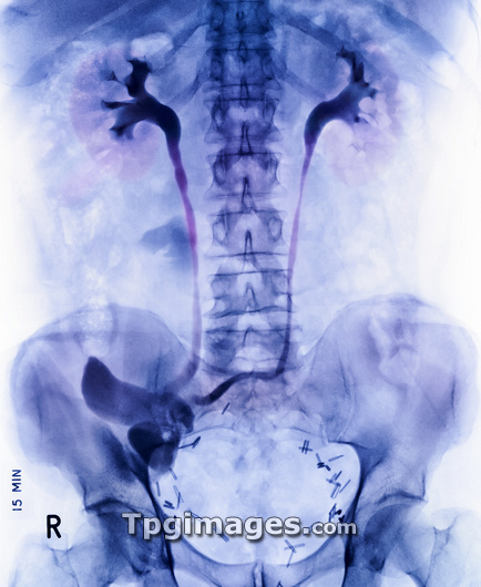
Ileal conduit surgery. Coloured frontal X-ray of a male patient shortly after having an ileal conduit urinary diversion. The spine runs down from top centre to the pelvis at lower centre. The kidneys are at upper left and right of the spine. An ileal conduit urinary diversion is performed when the urine produced by the kidneys (upper left and right) cannot continue as normal to the bladder. The surgery involves diverting the ureters (tubes connecting the kidneys and bladder) to a detached section of ileum (part of the small intestine, lower left). The end of the ileum section is then connected to an opening (stoma) in the abdominal wall, where the urine is collected by a disposable bag. The dark lines at lower left and right are surgical staples or stitches (sutures).
| px | px | dpi | = | cm | x | cm | = | MB |
Details
Creative#:
TOP03215348
Source:
達志影像
Authorization Type:
RM
Release Information:
須由TPG 完整授權
Model Release:
N/A
Property Release:
N/A
Right to Privacy:
No
Same folder images:
 Loading