
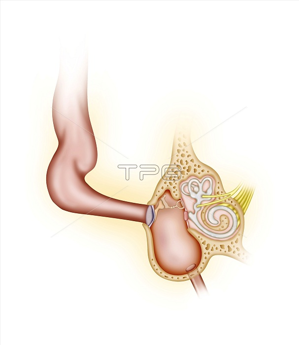
Dog ear anatomy. Illustration of the anatomy of a canine (dog) ear. The outer ear of a dog (upper left) is different to that of a human, being larger and more flexible, able to gather much more sound. The middle and inner ear (lower right) are the same as other mammals, with the sounds reaching the ear drum (tympanic membrane), and energy being transferred to the ear bones (ossicles: malleus, incus and stapes), and from there to the oval window of the vestibule of the inner ear. The sounds travel through the vestibular fluid and reach the cochlea (lower right, spiral shaped). This triggers responses that are transmitted to the brain via the auditory nerves (yellow). At bottom right is the Eustachian tube. The semicircular canals are above the cochlea.
| px | px | dpi | = | cm | x | cm | = | MB |
Details
Creative#:
TOP17480872
Source:
達志影像
Authorization Type:
RM
Release Information:
須由TPG 完整授權
Model Release:
N/A
Property Release:
N/A
Right to Privacy:
No
Same folder images:
 Loading