
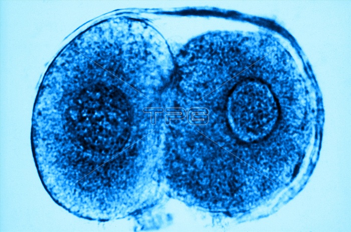
Color enhanced transmission electron micrograph of a human ovum, zygote cell stage, about 60 hours after fertilization. Immediately following the separation of chromosomes, the cell cleaves and becomes two cells called blastomeres. A nucleus can be seen in each. This occurs when a zygote undergoes its first in a series of mitotic divisions. Divisions will continue as the blastomeres move down the fallopian tubes to the uterine cavity. Magnification: x1000.
| px | px | dpi | = | cm | x | cm | = | MB |
Details
Creative#:
TOP22225828
Source:
達志影像
Authorization Type:
RM
Release Information:
須由TPG 完整授權
Model Release:
N/A
Property Release:
No
Right to Privacy:
No
Same folder images:
enhancementcolorizedtemtransmissionelectronmicroscopytransmissionelectronmicrographemelectronmicroscopyelectronmicrographmicroscopymicroscopicmicrographsciencenon-pathologicalnormalmedicalhealthyphysiologicalphysiologyhistologicalhistologycytologybiologicalbiologynucleusfertilizationfertilizeddevelopmentdevelopingdivisiondividingcelldivisioncellstagecelleukaryoticeukaryotemammalianreproductionreproductionhumanreproductionreproductivesystemhumanembryogenesisembryologyembryoidembryohumanembryoreproductivecellfemaleeggeggcellgameteovumhumanovumblastomeresdiploidcellzygote
biologicalbiologyblastomerescellcellcellcellcellcellcolorizedcytologydevelopingdevelopmentdiploiddividingdivisiondivisioneggeggelectronelectronelectronelectronemembryoembryoembryogenesisembryoidembryologyenhancementeukaryoteeukaryoticfemalefertilizationfertilizedgametehealthyhistologicalhistologyhumanhumanhumanhumanmammalianmedicalmicrographmicrographmicrographmicroscopicmicroscopymicroscopymicroscopynon-pathologicalnormalnucleusovumovumphysiologicalphysiologyreproductionreproductionreproductionreproductivereproductivesciencestagesystemtemtransmissiontransmissionzygote
 Loading