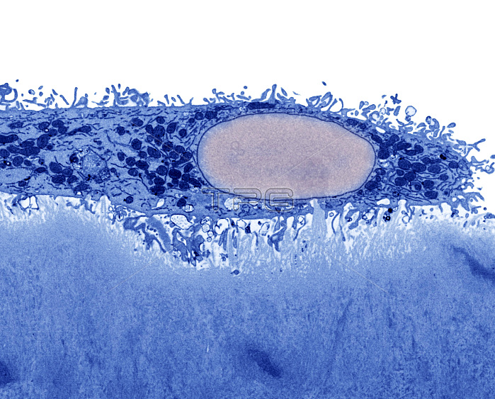
Transmission electron micrograph of a bone cell responsible for breaking down bone tissue (osteoclast; top half of image) forming a pit in dentine (dense calcified tissue that makes up the core of a tooth below the outer enamel; bottom half of image). A specialized cell membrane (ruffled border) forms between the surface of the osteoclast and the dentine. The nucleus of the osteoclast is also visible (pink). Horizontal image width is 41 microns. Supplied by Kevin Mackenzie, courtesy of Wellcome Images.
| px | px | dpi | = | cm | x | cm | = | MB |
Details
Creative#:
TOP22238977
Source:
達志影像
Authorization Type:
RM
Release Information:
須由TPG 完整授權
Model Release:
N/A
Property Release:
No
Right to Privacy:
No
Same folder images:

 Loading