
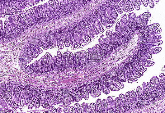
Small intestine, light micrograph. The inner epithelial lining of the intestine shows many finger-like processes called villi which result in a large surface area facing the lumen of the gut. This interface permits absorptive and secretory functions across the epithelium. The villi and the crypts between them belong to the intestinal mucosa which itself is thrown into major folds called plicae circulares that are visible macroscopically. The core of plicae is comprised of connective tissue with neurovascular structures and smooth muscle. Paraffin section, haematoxylin and eosin stain. Magnification: x30 when printed at 10cm wide.
| px | px | dpi | = | cm | x | cm | = | MB |
Details
Creative#:
TOP28610427
Source:
達志影像
Authorization Type:
RM
Release Information:
須由TPG 完整授權
Model Release:
n/a
Property Release:
n/a
Right to Privacy:
No
Same folder images:
 Loading