
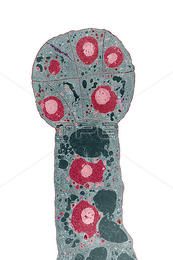
Coloured transmission electron micrograph (TEM) of a developing embryo of a turnip (Brassica campestris). This is a thin section of a young embryo, three days after pollination took place. The large light and dark red bodies are cell nuclei. The dark green empty spaces are vacuoles containing liquid, and the small coloured particles are organelles within the cytoplasm, including mitochondria and plastids. The embryo is globular; seen here as the circular group of small, dense cells, to top. The two larger cells below are part of a stalk called the suspensor. The embryo here consists of sixteen cells; seven are partly visible. As it grows, it will develop a clear polar axis, with a shoot apex between two cotyledons above, and a root tip to its base. The embryo here is isolated from the plant; in life it is bathed and fed by nutrients within the embryo sac. Magnification: x3000 at 10x8.
| px | px | dpi | = | cm | x | cm | = | MB |
Details
Creative#:
TOP26437437
Source:
達志影像
Authorization Type:
RM
Release Information:
須由TPG 完整授權
Model Release:
N/A
Property Release:
N/A
Right to Privacy:
No
Same folder images:
 Loading