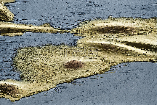
Coloured scanning electron micrograph (SEM) of cultured Ptk2 cells growing as a monolayer on glass. There are five cells in a diagonal line from bottom left, each with a raised surface feature corresponding to the position of the cell nucleus. The edges of the cells are thin and show finger-like extensions of the cell membrane. Ptk2 is a cell line derived from epithelial kidney tissue of a male long-nosed potoroo (Potorous tridactylis), a marsupial native to South Eastern Australia and Tasmania. The cells are used as a model for the study of nuclear division (mitosis). They contain only a small number of chromosomes (5 autosomes and 3 sex chromosomes), and retain their flattened shape, shown here, even during the cell division process. This enables direct observation of mitosis in living material by light microscopy, avoiding the preparative techniques of electron microscopy. Magnification: x600 at 10x8.
| px | px | dpi | = | cm | x | cm | = | MB |
Details
Creative#:
TOP26437450
Source:
達志影像
Authorization Type:
RM
Release Information:
須由TPG 完整授權
Model Release:
N/A
Property Release:
N/A
Right to Privacy:
No
Same folder images:

 Loading