
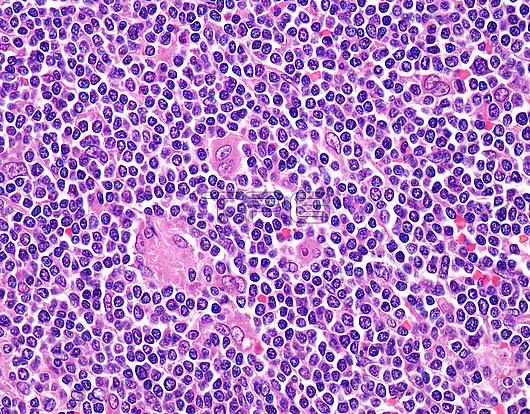
Mantle cell lymphoma, light micrograph. Mantle cell lymphoma (MCL) is an aggressive B-cell lymphoma that makes up about 2.5% of non-Hodgkin lymphomas. It usually presents at advanced stages in the 5th and 6th decades of life with generalized lymphadenopathy and bone marrow involvement. There may also be involvement of peripheral blood, spleen, liver, gastrointestinal tract (lymphomatoid polyposis), Waldeyer's ring, lung, and pleura. This image shows mantle cell lymphoma presenting as lymphomatous polyposis coli in a middle-aged male. The patient presented with diarrhoea, bleeding per rectum and weight loss. Colonoscopy showed multiple polyps in the colon. Colonic biopsy (shown here) revealed diffuse sheets and nodular aggregates of medium-sized atypical lymphoid cells with irregular, indented nuclei and punctate nucleoli. A few larger atypical lymphoid cells with more dispersed chromatin and prominent nucleoli were also present. Mitotic activity was increased. The diagnosis was confirmed on morphology and immunohistochemical profile. Cytogenetics showed t(11;14)(q13;q32) translocation.
| px | px | dpi | = | cm | x | cm | = | MB |
Details
Creative#:
TOP28646153
Source:
達志影像
Authorization Type:
RM
Release Information:
須由TPG 完整授權
Model Release:
n/a
Property Release:
n/a
Right to Privacy:
No
Same folder images:
 Loading