
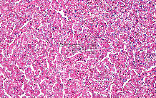
Light micrograph of necrotic tumour cells. Often, tumours which outgrow their blood have areas that are necrotic or dead. Necrosis can be identified histologically by loss of nuclei of the necrotic cells, leaving behind only pink cytoplasm. Another feature of necrosis is karyorrhectic debris (very small blue dots), which are the remnants of the nuclei that have been broken down by the process of the cells??death. Haematoxylin and eosin stained tissue section. Magnification: 200x when printed at 10cm.
| px | px | dpi | = | cm | x | cm | = | MB |
Details
Creative#:
TOP29676305
Source:
達志影像
Authorization Type:
RM
Release Information:
須由TPG 完整授權
Model Release:
N/A
Property Release:
N/A
Right to Privacy:
No
Same folder images:
pathologypathologicalmedicinemedicaldiseasedisorderconditionabnormalunhealthyanatomicpathologynecrosistumornecrosismalignancycanceroncologysurgicalpathologyhistopathologicalhistopathologymicroscopylmlightmicrographslidehematoxylinandeosinhaematoxylinandeosinhumanbodyanatomytissuecellsdiseasenobodyno-one
abnormalanatomicanatomyandandbodycancercellsconditiondiseasediseasedisordereosineosinhaematoxylinhematoxylinhistopathologicalhistopathologyhumanlightlmmalignancymedicalmedicinemicrographmicroscopynecrosisnecrosisno-onenobodyoncologypathologicalpathologypathologypathologyslidesurgicaltissuetumorunhealthy
 Loading