
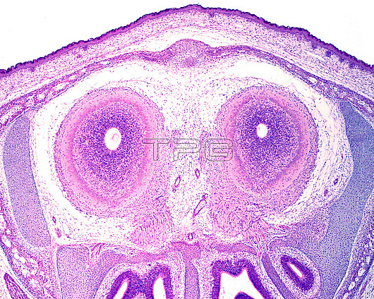
Light micrograph of the cross-sectioned olfactory bulb of a rat embryo. It shows a small central lumen surrounded by a ventricular proliferating layer. The mitral cell and outer plexiform layers can already be recognised, as well as the olfactory nerve fibres directed toward the nasal cavity.
| px | px | dpi | = | cm | x | cm | = | MB |
Details
Creative#:
TOP29728571
Source:
達志影像
Authorization Type:
RM
Release Information:
須由TPG 完整授權
Model Release:
N/A
Property Release:
N/A
Right to Privacy:
No
Same folder images:
olfactorybulbnasalcavitydevelopmentmitralcellembryoembryoniceosinhaematoxylinhematoxylinhistologicalhistologylightmicroscopemicroscopicalmicroscopynasalolfactoryturbinateslightmicrographlmmicroscopynobodyno-onehistologyhistologicalhealthynormalanimaldevelopmentalbiologyembryologyembryologicalbiologicalsectionsectioned
animalbiologicalbiologybulbcavitycelldevelopmentdevelopmentalembryoembryologicalembryologyembryoniceosinhaematoxylinhealthyhematoxylinhistologicalhistologicalhistologyhistologylightlightlmmicrographmicroscopemicroscopicalmicroscopymicroscopymitralnasalnasalno-onenobodynormalolfactoryolfactorysectionsectionedturbinates
 Loading