
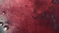
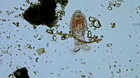
Mitosis cell division. Labelled animation showing the stages of mitotic cell division (mitosis). At the start of the clip, the chromosomal genetic material (red and blue) is shown in the nucleus (orange). Within the nucleus is the nucleolus (round). The nucleus is bounded by the nuclear membrane. Outside the nucleus, structures called centrioles (black, centre left) are shown. During mitotic cell division, the chromosomes replicate and the nuclear membrane dissolves. The centrioles move to opposite ends of the cell and the chromosomes line up down the centre of the cell. Microtubules form from the centrioles towards the chromosomes, creating what is called the mitotic spindle. The spindle fibres attach to the chromosomes and pull them apart. The cell divides into two, with a set of identical chromosomes in each daughter cell. The spindle fibres dissolve and the nuclear membrane reforms with a new nucleolus and nucleus in each cell. The stages are named prophase, metaphase, anaphase, telophase and cytokinesis. For this animation without labels, see clip K004/6133.


Details
WebID:
C01826956
Clip Type:
RM
Super High Res Size:
1920X1080
Duration:
00:44.0
Format:
QuickTime
Bit Rate:
25 fps
Available:
download
Comp:
200X112 (0.00 M)
Model Release:
NO
Property Release
No
 Loading