
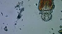
Slipped lumbar disc. Sequence of magnetic resonance imaging (MRI) axial scans showing the internal structure in the lower back of a 30-year-old woman with a prolapsed (slipped) intervertebral disc. The front of the body is at top in this view from below, with the rear of the body and the spine at lower centre. The sequence moves vertically along the spine from bottom to top. The slipped disc, which is at the L4/L5 level (see K003/7757), is seen early on in the sequence. It is indenting the thecal sac of the cauda equina (a bundle of spinal nerves) and is compressing the nerve roots of the cauda equina. This has caused severe and sudden (acute) back pain, loss of bladder sensation, and urinary incontinence.


Details
WebID:
C01843303
Clip Type:
RM
Super High Res Size:
1920X1080
Duration:
00:00:04.000
Format:
QuickTime
Bit Rate:
24 fps
Available:
download
Comp:
200X150 (0.00 M)
Model Release:
NO
Property Release
No
 Loading