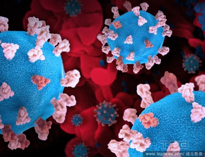
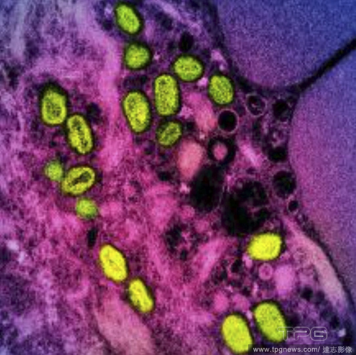
EditorialAn undated image provided by the National Institute of Allergy and Infectious Diseases shows a colorized transmission electron micrograph of monkeypox particles (green) found within an infected cell (pink and purple), cultured in the laboratory. (National Institute of Allergy and Infectious Diseases via The New York Times)
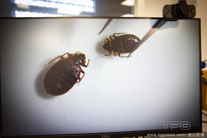
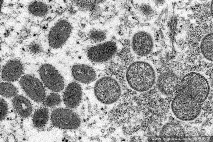
EditorialAn electron microscope image provided by the U.S. Centers for Disease Control and Prevention shows oval-shaped, mature monkeybpox virions, left, and spherical, immature monkeypox virions, right, in 2003. (Centers for Disease Control and Prevention via The New York Times)


 Loading