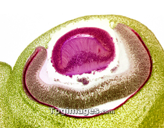
Developing pig eye. Coloured light micrograph of the eye of a 20mm pig embryo (Sus scrofa domestica), showing the lens (pink), the retina (brown), the choroid (dark pink line) and the sclera (white of the eye, green). The eye works by allowing light to be focused by the lens onto the retina. The retina contains photoreceptor cells, which allow the eye to distinguish between colours (cone cells) and to see at night (rod cells). The choroid layer lines the inside of the eye underneath the retina and is pigmented to prevent light reflecting inside the eye and distorting the image. Magnification: x180 when printed 10cm wide.
| px | px | dpi | = | cm | x | cm | = | MB |
Details
Creative#:
TOP03222249
Source:
達志影像
Authorization Type:
RM
Release Information:
須由TPG 完整授權
Model Release:
N/A
Property Release:
N/A
Right to Privacy:
No
Same folder images:

 Loading