
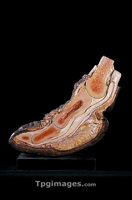
19th century model of a camel's toe. Coloured plaster moulding of a sagittal section through the toe of a dromedary camel (Camelus dromedarius), showing the structure of the bones, tendons and muscles. This anatomical model was made by Andre Richir in about 1950 as a veterinary teaching aid. Photographed in the museum of the National Veterinary School of Alfort, Maisons-Alfort, France.
| px | px | dpi | = | cm | x | cm | = | MB |
Details
Creative#:
TOP06660379
Source:
達志影像
Authorization Type:
RM
Release Information:
須由TPG 完整授權
Model Release:
NO
Property Release:
NO
Right to Privacy:
No
Same folder images:
dromedarycamelcamelusdromedariusanimalequipmentmammaleuropemaisons-alfortfranceanatomybiologyhistoryzoologyone120thcentury1950anatomicalandrerichiranimalsbiologicalblackbackgroundbonebonescamelcutoutcutoutscut-outcut-outscutoutcutoutsecolenationaleveterinaired'alforteducationeducationaleuropeanfaunafootfrenchhistoricalmetatarsalsmetatarsusmodelmouldmouldingmusclestructuremusclesmuseummyologicalmyologynationalveterinaryschoolofalfortnatureplastersagittalsectionsectionedsingleteachingaidtendontendonstoevetveterinarymedicineveterinarysciencewildlifezoological"
"1195020thaidalfortanatomicalanatomyandreanimalanimalsbackgroundbiologicalbiologyblackbonebonescamelcamelcameluscenturycutcutcut-outcut-outscutoutcutoutsd'alfortdromedariusdromedaryecoleeducationeducationalequipmenteuropeeuropeanfaunafootfrancefrenchhistoricalhistorymaisons-alfortmammalmedicinemetatarsalsmetatarsusmodelmouldmouldingmusclemusclesmuseummyologicalmyologynationalnationalenatureofoneoutoutsplasterrichirsagittalschoolsciencesectionsectionedsinglestructureteachingtendontendonstoevetveterinaireveterinaryveterinaryveterinarywildlifezoologicalzoology
 Loading