
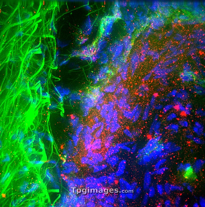
Oesophagus nerves. Fluorescence deconvolution micrograph of a section through a healthy oesophagus (gullet), showing the adventitial nerves (green, left) of the outer wall. The different colours show different components; g-actin: red, smooth muscle actin: blue. Magnification x40 when printed 10 centimetres wide.
| px | px | dpi | = | cm | x | cm | = | MB |
Details
Creative#:
TOP06663265
Source:
達志影像
Authorization Type:
RM
Release Information:
須由TPG 完整授權
Model Release:
NO
Property Release:
NO
Right to Privacy:
No
Same folder images:
humanhumanbodyanatomybiologyhistologymedicinelightmicrographfluorescentdeconvolutionmicrographconfocallightmicrographlightmicroscopeactinadventitialnerveanatomicalbiologicalcellcellsesophagealesophagusfluorescenceg-actingastrointestinalgullethealthyhistologicalhistopathologicallmmedicalmusclesmuscularnervesnervoussystemnormaloesophagealoesophagusouteroesophaguspeoplepersonproteinsectionsectionedsmoothmusclestructuretissuewall"
"actinadventitialanatomicalanatomybiologicalbiologybodycellcellsconfocaldeconvolutionesophagealesophagusfluorescencefluorescentg-actingastrointestinalgullethealthyhistologicalhistologyhistopathologicalhumanhumanlightlightlightlmmedicalmedicinemicrographmicrographmicrographmicroscopemusclemusclesmuscularnervenervesnervousnormaloesophagealoesophagusoesophagusouterpeoplepersonproteinsectionsectionedsmoothstructuresystemtissuewall
 Loading