
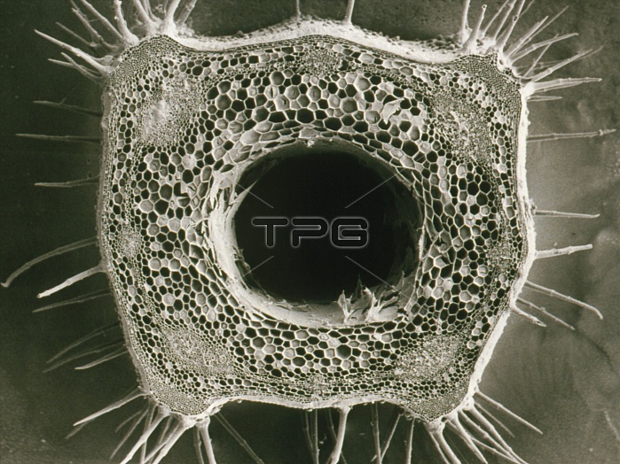
Scanning electron micrograph of a cut stem of the white dead nettle, Lamium album. The hollow centre is produced by the collapse of thin-walled pith cells. Surrounding this core is a layer of unspec- ialised cortical cells - the cortical parenchyma. Eight vascular bundles serve to conduct water & nutrients: one near each of the 4 corners, & one half way along each side of the stem. In the extreme corners of the stem, the best position mechanically, small collenchyma cells are visible. The spikes on the outside of the stem are hairs, designed to discourage insects from climbing the plant. Magnification: x6 at 35mm size, x40 at 8x10- inch size. Reference: MICROCOSMOS, figure 4.6, page 70.
| px | px | dpi | = | cm | x | cm | = | MB |
Details
Creative#:
TOP10173588
Source:
達志影像
Authorization Type:
RM
Release Information:
須由TPG 完整授權
Model Release:
N/A
Property Release:
N/A
Right to Privacy:
No
Same folder images:
 Loading