
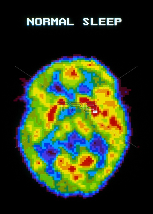
Brain during normal sleep. Coloured Positron Emission Tomography (PET) scan of the human brain during normal sleep. Colour-coding depicts active cerebral brain areas (red) through to inactive areas (blue). During normal sleep the brain is slightly active, less active than when awake, but more active than in deep sleep. Brain activity increases further during REM (rapid eye movement) dreaming" sleep. PET scanning shows metabolic activity of the brain. A radioactive tracer (here, radio-labelled glucose) is injected into the blood and absorbed by active tissues of the brain. The PET scanner detects photons emitted by the tracer, to produce a "slice" image of the brain."
| px | px | dpi | = | cm | x | cm | = | MB |
Details
Creative#:
TOP10214974
Source:
達志影像
Authorization Type:
RM
Release Information:
須由TPG 完整授權
Model Release:
N/A
Property Release:
N/A
Right to Privacy:
No
Same folder images:
 Loading