
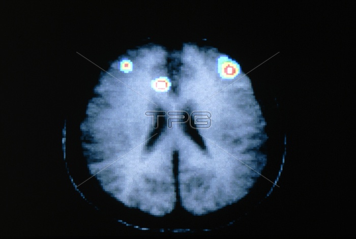
Memory processing. Coloured positron emission tomography (PET) scan of the brain during memory processing. The PET scan has been superimposed onto a grey magnetic resonance imaging (MRI) scan. In this axial (horizontal) section the front of the brain is at top, with ventricles (cavities, black) at centre. The scan shows brain activity associated with memory processing occurring in three areas (red & white) of the prefrontal cortex. PET scans use radioactively-labelled substances introduced into the blood to view metabolic activity.
| px | px | dpi | = | cm | x | cm | = | MB |
Details
Creative#:
TOP10219334
Source:
達志影像
Authorization Type:
RM
Release Information:
須由TPG 完整授權
Model Release:
N/A
Property Release:
N/A
Right to Privacy:
No
Same folder images:
 Loading