
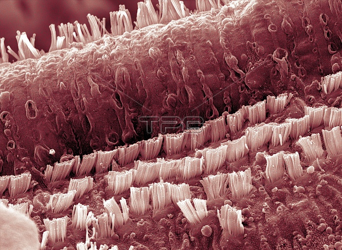
Cochlea cells. Coloured scanning electron micrograph (SEM) of hair cells in the cochlea of a human ear. The rows of columnar outer pillar cells run along the organ of Corti, the auditory sense organ. The outer pillar cells arise from the basilar membrane, and their upper surfaces form part of the surface of the organ of Corti. This organ lies on the basilar membrane, an internal surface of the cochlear duct. The tectorial membrane, which overlies the sensory hairs, has been removed. Sound waves deform hair cell cilia & trigger auditory nerve impulses. Magnification: x5000 at 6x7cm size.
| px | px | dpi | = | cm | x | cm | = | MB |
Details
Creative#:
TOP10220196
Source:
達志影像
Authorization Type:
RM
Release Information:
須由TPG 完整授權
Model Release:
N/A
Property Release:
N/A
Right to Privacy:
No
Same folder images:
 Loading