
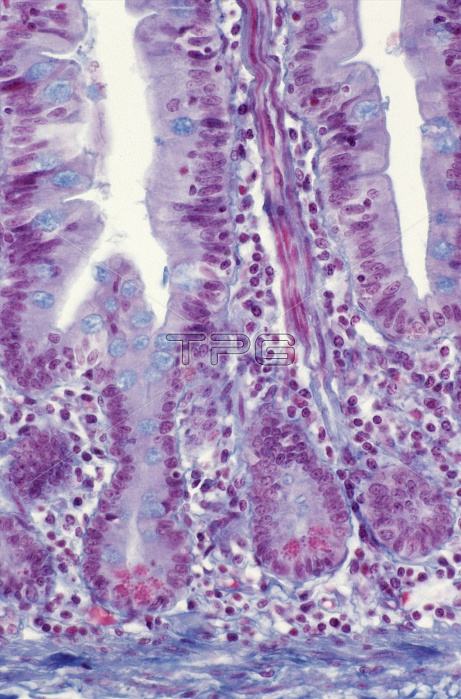
Small intestine lining. Light micrograph of a section through the lining (mucosa) of the small intestine. It shows crypts of Lieberkuhn (purple folds, lower) between the intestinal villi (projections, upper). The crypts produce new cells by constant cell division; these progress up the villi to renew the intestinal lining. The lining of the villi and crypts is composed of columnar enterocyte cells, which are involved in digestion and absorption, and mucus-secreting goblet cells (blue). At the base of the crypts are Paneth cells (stained red), which are thought to protect the intestine from disease. At bottom is a muscle layer (blue). Magnification unknown.
| px | px | dpi | = | cm | x | cm | = | MB |
Details
Creative#:
TOP10220624
Source:
達志影像
Authorization Type:
RM
Release Information:
須由TPG 完整授權
Model Release:
N/A
Property Release:
N/A
Right to Privacy:
No
Same folder images:
 Loading