
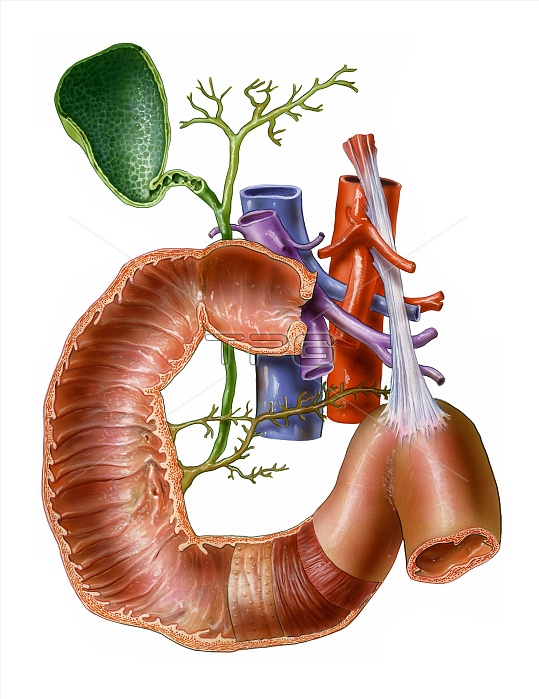
Small intestine, artwork. At centre is the pylorus, the point where the stomach (not seen) opens into the duodenum, the first part of the small intestine. The duodenal wall contains plicae, small folds, which are covered in small projections called villi. These increase the surface area for digestion and absorption of food. At bottom centre are the layers of the duodenum wall, the submucosa, which contains blood vessels, circular muscles and longitudinal muscles. At centre is the portal vein (purple, which serves the liver), the vena cava (blue, the body's main vein) and the aorta (red, the body's main artery). The gall bladder (green) and pancreatic ducts (yellow) are also seen.
| px | px | dpi | = | cm | x | cm | = | MB |
Details
Creative#:
TOP10220659
Source:
達志影像
Authorization Type:
RM
Release Information:
須由TPG 完整授權
Model Release:
N/A
Property Release:
N/A
Right to Privacy:
No
Same folder images:
 Loading