
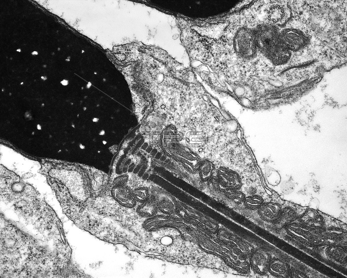
Human spermatid. Transmission electron micrograph (TEM) of a section through an elongating spermatid in the human testis (testicle), showing the densely compacted nucleus (black, upper left) and part of the tail (flagellum, centre to bottom right). Spermatids mature into spermatozoa (sperm cells). The flagellum is anchored to the spermatid nucleus by striated protein segments. Extending from this are two long columns (black lines) of proteins comprising part of the nine outer dense fibres of the flagellum. Mitochondria form coiled helices around the tail extending as far as the middle piece of the flagellum, and supply the energy needed for sperm motility. Magnification: x70,000, when printed 10 centimetres wide.
| px | px | dpi | = | cm | x | cm | = | MB |
Details
Creative#:
TOP10393045
Source:
達志影像
Authorization Type:
RM
Release Information:
須由TPG 完整授權
Model Release:
No
Property Release:
No
Right to Privacy:
No
Same folder images:
 Loading