
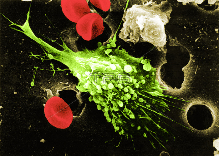
Step one in a six-step sequence of the death of a cancer cell. A cancer cell (green) has migrated through the holes of a matrix coated membrane from the top to the bottom, simulating natural migration of an invading cancer cell between, and sometimes through, the vascular endothelium. Notice the spikes or pseudopodia that are characteristic of an invading cancer cell (1). A buffy coat containing red blood cells, lymphocytes and macrophages is added to the bottom of the membrane. A group of macrophages identify the cancer cell as foreign matter and start to stick to the cancer cell, which still has its spikes (2). Macrophages begin to fuse with, and inject its toxins into, the cancer cell. The cell starts rounding up and loses its spikes (3). As the macrophage cell becomes smooth (4). The cancer cell appears lumpy in the last stage before it dies. These lumps are actually the macrophages fused within the cancer cell (5). The cancer cell then loses its morphology, shrinks up and dies (6). Photo magnification: 1: x12,000; 2: x4,000; 3: x8,000; 4: x26,000; 5: x56,000; 6: x14,000. Full sequence: BJ4579, BS9471, BS9472, BS9473, BS9474, BS9475.
| px | px | dpi | = | cm | x | cm | = | MB |
Details
Creative#:
TOP22229172
Source:
達志影像
Authorization Type:
RM
Release Information:
須由TPG 完整授權
Model Release:
N/A
Property Release:
No
Right to Privacy:
No
Same folder images:
 Loading