
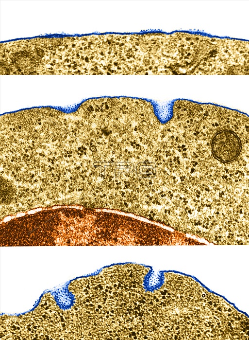
Color enhanced transmission electron micrographs of portions of the surface of polychromatophilic erythroblasts from guinea pig bone marrow. This series of images illustrates the intermediate stages in the formation of coated micropinocytotic vesicles. We see small thickened areas of membrane acquire an inner coat and a fuzzy external layer to which particles of ferritin adhere.
| px | px | dpi | = | cm | x | cm | = | MB |
Details
Creative#:
TOP22229943
Source:
達志影像
Authorization Type:
RM
Release Information:
須由TPG 完整授權
Model Release:
N/A
Property Release:
No
Right to Privacy:
No
Same folder images:
 Loading