
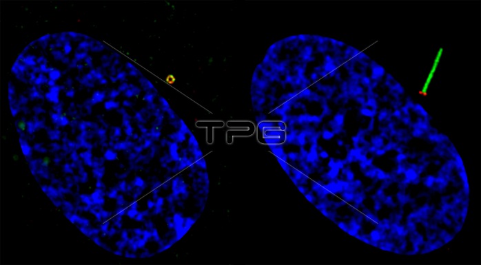
Defects in ciliary formation and function are linked to several human diseases, including cancer. Some cancer cells appear to have lost the ability to develop or maintain cilia, which may contribute to tumorigensis. Here, structured illumination microscopy imaging of RPE cells shows ciliogenesis progression. Ciliogenesis starts from docking pre-ciliary vesicles to the distal appendage of the mother centriole. Those docked vesicles fuse to form a larger ciliary vesicle and then extend to the ciliary membrane surrounding the growing microtubule-based axoneme. A GFP-labeled G protein-coupled receptor called Smoothened localizes to these docked vesicles and also the ciliary membrane. Left cell shows GFP-Smoothened (green) vesicles docked at the mother centriole/basal body distal appendage (marked by CEP164 in red). Right cell shows ciliary GFP-Smoothened localized on ciliary membrane. Nuclei (blue).
| px | px | dpi | = | cm | x | cm | = | MB |
Details
Creative#:
TOP22235322
Source:
達志影像
Authorization Type:
RM
Release Information:
須由TPG 完整授權
Model Release:
N/A
Property Release:
No
Right to Privacy:
No
Same folder images:
 Loading