
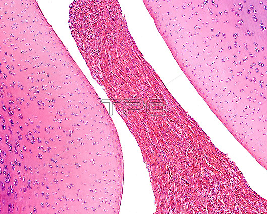
Light micrograph of a longitudinal section of a knee of a young experimental animal. The epiphysis of the femur (above) is made up of hyaline cartilage and the epiphyseal ossification nucleus has not yet formed. However, in the tibial epiphysis (bottom) the epiphyseal ossification nucleus and the metaphyseal cartilage are evident. Between both articular surfaces there is a narrow articular cavity. At right is the patella and a small wedge of fibrous connective tissue that penetrates the joint cavity corresponding to the meniscus. Below the patella is the attachment of the joint capsule to the periosteum of the tibia.
| px | px | dpi | = | cm | x | cm | = | MB |
Details
Creative#:
TOP26624882
Source:
達志影像
Authorization Type:
RM
Release Information:
須由TPG 完整授權
Model Release:
N/A
Property Release:
N/A
Right to Privacy:
No
Same folder images:
 Loading