
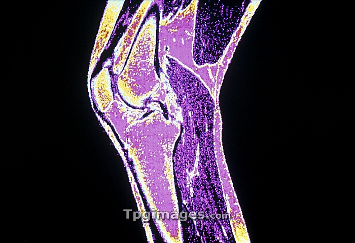
Knee joint. Coloured Magnetic Resonance Image (MRI) of a sagittal section through a human knee joint. The knee is the joint between the femur or thigh bone (top, pink and yellow) and tibia bone (bottom, pink). The articulation surfaces of these bones are covered by cartilage; the bones are joined by two cruciate ligaments. The joint cavity is filled with a synovial fluid, which lubricates the movement of the joint. MRI scanning uses radio waves and an electromagnet to create \slice\" images through the body."
| px | px | dpi | = | cm | x | cm | = | MB |
Details
Creative#:
TPG05325323
Source:
達志影像
Authorization Type:
RF
Release Information:
須由TPG 完整授權
Model Release:
NO
Property Release:
NO
Right to Privacy:
No
Same folder images:
 Loading