
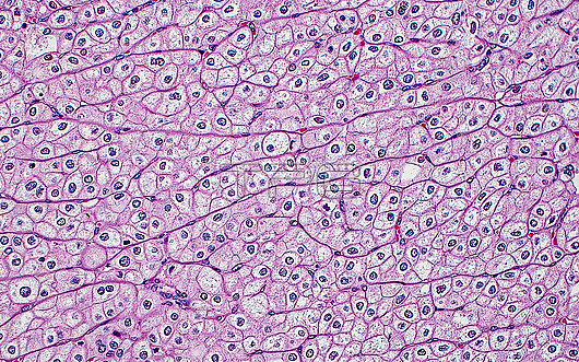
Light micrograph of chromophobe renal cell carcinoma. Distinctive histopathologic features of chromophobe carcinoma that can be seen in this image include distinct ?œplant-like??cell membranes, clear perinuclear halos, and binucleation (presence of two nuclei in a single cell). Haematoxylin and eosin stained tissue section. Magnification: 200x when printed at 10cm.
| px | px | dpi | = | cm | x | cm | = | MB |
Details
Creative#:
TOP29676289
Source:
達志影像
Authorization Type:
RM
Release Information:
須由TPG 完整授權
Model Release:
N/A
Property Release:
N/A
Right to Privacy:
No
Same folder images:
pathologypathologicalmedicinemedicaldiseasedisorderconditionabnormalunhealthyanatomicpathologykidneykidneycancerrenalrenalcancerrenalcellcarcinomachromophobechromophoberenalcellcarcinomacanceroncologyurologynephrologygenitourinarygenitourinarypathologyhistopathologicalhistopathologymicroscopylmlightmicrographslidehematoxylinandeosinhaematoxylinandeosinhumanbodyanatomytissuecellsnobodyno-one
abnormalanatomicanatomyandandbodycancercancercancercarcinomacarcinomacellcellcellschromophobechromophobeconditiondiseasedisordereosineosingenitourinarygenitourinaryhaematoxylinhematoxylinhistopathologicalhistopathologyhumankidneykidneylightlmmedicalmedicinemicrographmicroscopynephrologyno-onenobodyoncologypathologicalpathologypathologypathologyrenalrenalrenalrenalslidetissueunhealthyurology
 Loading