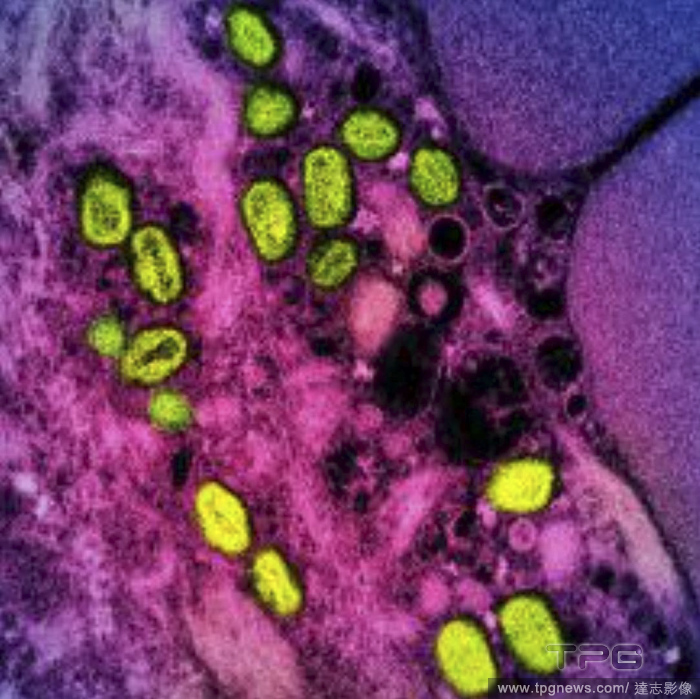
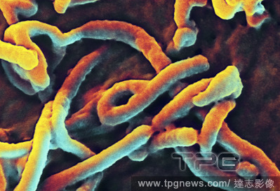
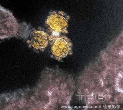
EditorialAn undated image provided by the National Institute of Allergy and Infectious Diseases shows a colorized transmission electron micrograph of monkeypox particles (green) found within an infected cell (pink and purple), cultured in the laboratory. (National Institute of Allergy and Infectious Diseases via The New York Times)
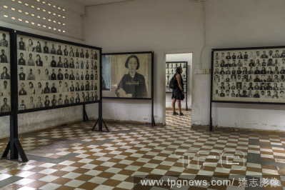
EditorialPhotographs of victims of the Khmer Rouge at the Tuol Sleng Genocide Museum in Phnom Penh, Cambodia, on Nov. 15, 2018. (Adam Dean/The New York Times)
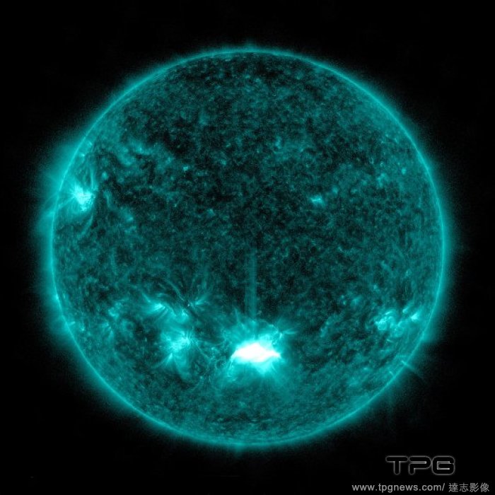
EditorialA colorized mosaic of the surface of Venus, seen through its clouds by the Magellan spacecraft in the early 1990s. (USGS Astrogeology Science Center via The New York Times)
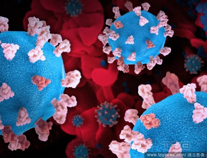
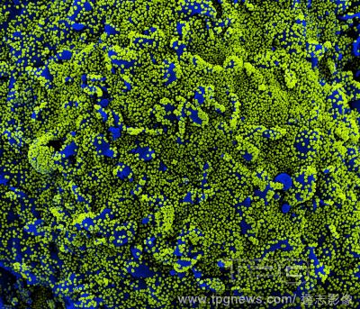
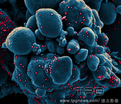
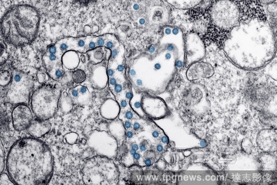
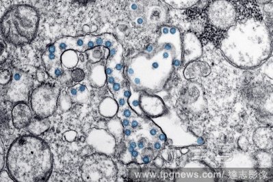
EditorialA colorized scanning electron micrograph of a cell (blue) heavily infected with coronavirus particles isolated from a patient sample in December 2020. (NIAID/National Institutes of Health via The New York Times)
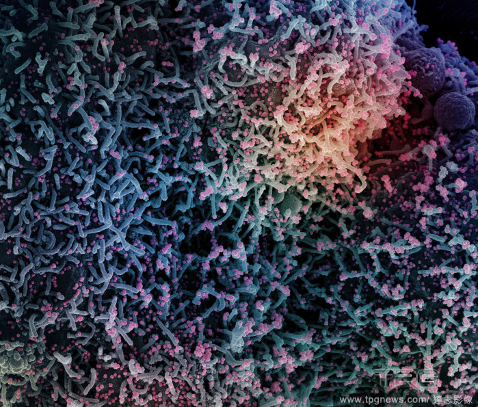
EditorialIn a photo from the National Institute of Allergy and Infectious Diseases, a colorized scanning electron micrograph of a dying cell infected with the coronavirus, with virus particles in red. (National Institute of Allergy and Infectious Diseases via The New York Times)
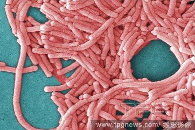
EditorialA digitally colorized scanning electron microscopic (SEM) image provided by Janice Haney Carr/Centers for Disease Control and Prevention shows Legionella pneumophila, the bacteria that causes Legionnaires’ Disease. (Janice Haney Carr/Centers for Disease Control and Prevention via The New York Times)


 Loading